Chronic venous disease of the lower limbs, the condition behind so-called varicose veins, is one of the most frequent diseases in our population: it affects up to 56% of men and 60% of women (Epidemiology of chronic venous disease. Phlebology. 2008;23(3):103-11).
Up to 2% of the population reaches the most severe stages of the disease, developing chronic skin ulceration linked to venous hypertension (Ma H. The real cost of treating venous ulcers in a contemporary vascular practice. J Vasc Surg Venous Lymphat Disord. 2014 Oct;2(4):355-61).
Diagnosing chronic venous disease requires a detailed review of the patient's clinical history and an accurate description of signs and symptoms. The most common symptoms are aching, cramps, heaviness, fatigue, swelling, itching, tingling, heat and restless leg.
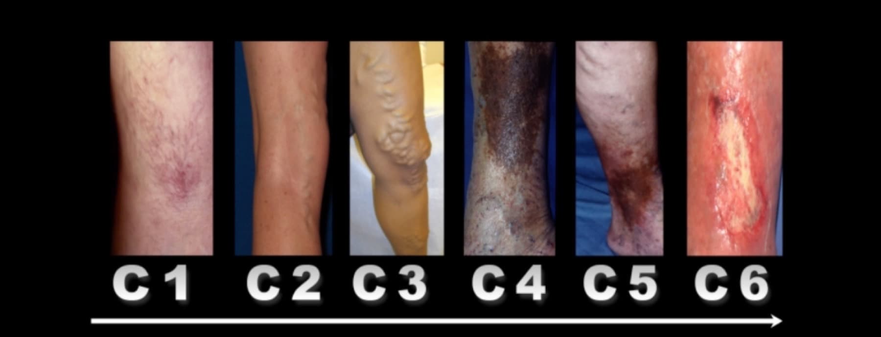
The leg can present six progressive steps of impaired venous drainage.
1. Spider veins
Small-calibre vessels such as telangiectasias (under 1 mm) and reticular veins (under 3 mm), affected by modest flow alterations and therefore visible and cosmetically unwelcome. In 26% of cases this clinical class is associated with alterations of the deeper venous system (Telangiectasia in the Edinburgh Vein Study: epidemiology and association with trunk varices and symptoms. Eur J Vasc Endovasc Surg 2008;36:719–724).
2. Varicose veins
Dilatation of the larger veins of the lower limbs. These vessels are usually tortuous and clearly visible, but they may also be invisible when they lie deep inside the leg. Contrary to common belief, these vessels are not the saphenous vein, but rather its “daughter” veins (CHIVA: instructions for users. Phlebology Journal 2014). In varicose disease these daughter veins, instead of carrying blood into the saphenous vein and from there towards heart and lungs, “steal” that blood from the saphenous vein and direct it towards the surface of the leg.
3. Varicose veins with oedema
The same venous dilatation as the previous stage, more advanced and associated with oedema. Oedema means an increase in the body fluid between cells, caused by impaired venous drainage from the lower limb towards the heart.
4. Lipodermatosclerosis
Rust-coloured skin pigmentation, usually around the ankle and foot and in the lower third of the leg. It arises from the increased pressure inside the veins affected by chronic insufficiency, and therefore from a reflux that pushes out the iron contained in the haemoglobin. That iron is deposited in the tissues and becomes, to all intents and purposes, a tattoo.
5. Healed ulcer
This stage describes cases in which a venous ulceration (an open wound on the leg caused by a venous problem) occurred and has now healed. Once a patient has had an ulceration, even a healed one, they belong for life to the fifth stage and therefore to the maximum severity of the disease: anyone who has had an ulcer keeps a significantly higher risk of having another one than someone who never had the problem. Specifically, up to 69% of patients with an ulcer will see the lesion return (What's new: management of venous leg ulcers: approach to leg ulcers. J Am Acad Dermatol 2016;74:627-40).
6. Open ulcer
The most severe stage, characterised by an open lesion of the skin. The ulceration is caused by the release of inflammatory products linked to the failure of venous return from the most peripheral parts of the leg towards the heart and lungs. The lesion can be small or large and can affect the patient's quality of life as much as chemotherapy (Ma H. The real cost of treating venous ulcers in a contemporary vascular practice. J Vasc Surg Venous Lymphat Disord. 2014 Oct;2(4):355-61).
The duplex ultrasound examination
Once the specialist has completed the detailed collection of clinical history, symptoms and signs of venous disease, an equally detailed duplex ultrasound examination has to be performed. It is an ultrasound scan, therefore non-invasive, to be carried out on both legs, regardless of whether the clinical signs are present on one limb only (Superficial vein thrombosis: a consensus statement. Int Angiol 2012;31:203-16).
This recommendation comes from the higher risk of developing venous problems in the leg opposite the one with evident disease (Leg symptoms of somatic, psychic, and unexplained origin in the population based Bonn Vein Study. Eur J Vasc Endovasc Surg 2013;46:255-62).
The examination must cover every venous compartment: superficial, saphenous and deep. If reflux is found in veins deeper than those visible to the naked eye, the risk of developing varicose disease increases 4.4 to 7.3 times over the following 13 years (Incidence and Risk Factors for Venous Reflux in the General Population: Edinburgh Vein Study 2014;48(2):208-214).
Why the operator matters
It is advisable that the same specialist who will perform any treatment also performs the detailed ultrasound examination (CHIVA: hemodynamic concept, strategy and results. International Angiology 2016;35(1):8-30). Unlike an X-ray or a CT scan, ultrasound is operator-dependent: the result depends on what the specialist chose to look at, and how they looked at it.
The correlation between the clinical picture reported by the patient, the visible signs of disease and the ultrasound findings is what defines the best treatment pathway for the venous condition: a pathway built today on several possible strategies, chosen according to the individual case.